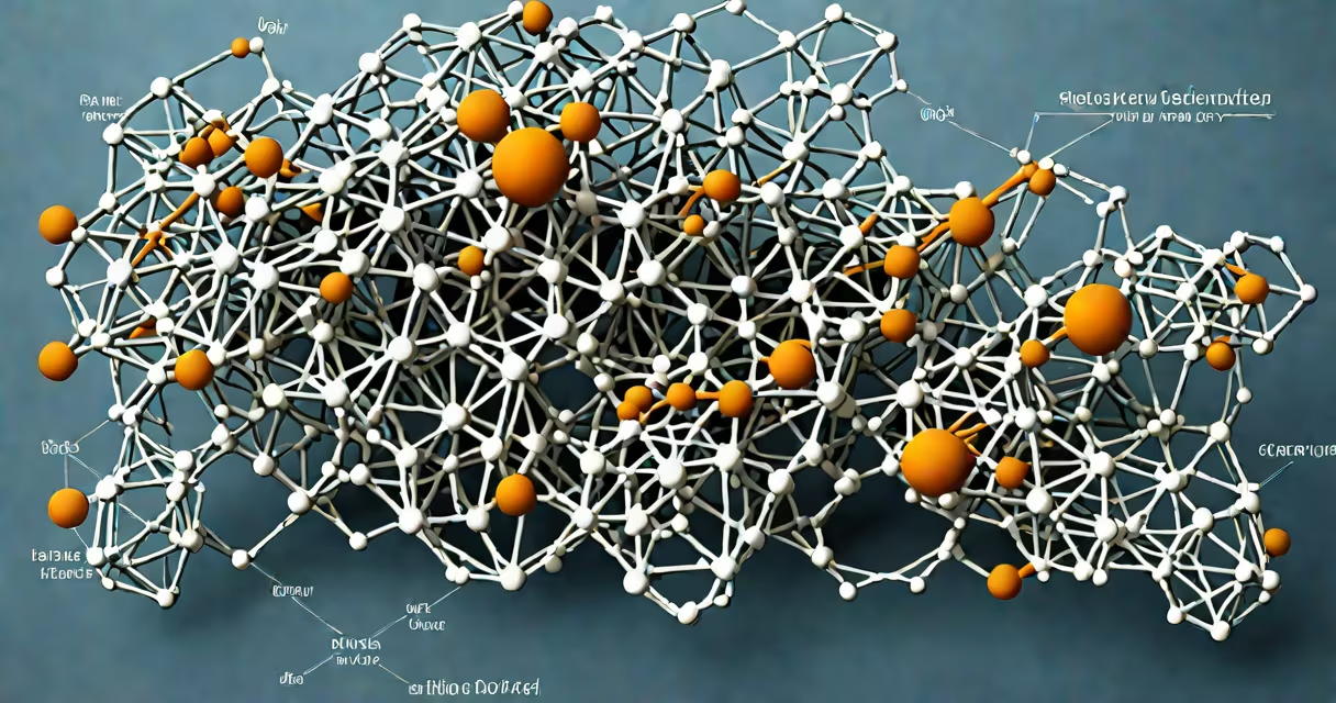
Amino acid residues are the individual units derived from amino acids after they form peptide bonds in a polypeptide chain. Each residue contributes its unique side chain to the structure and function of a protein.
When two amino acids join via a dehydration reaction, the carboxyl group of one links to the amino group of another, forming a covalent peptide bond. The resulting molecule loses a water molecule and becomes a single unit called a residue. This process repeats to build long chains known as polypeptides. Once folded into functional shapes, these chains become proteins.
Residues are not static-they influence each other through steric effects, hydrogen bonding, hydrophobic interactions, and electrostatic forces. These interactions govern how the chain folds into secondary structures like alpha helices and beta sheets, which then assemble into tertiary and quaternary structures. Without proper residue arrangement, proteins fail to function correctly.
Each amino acid begins as a free molecule with an amino group (-NH₂), a carboxyl group (-COOH), and a variable side chain (R-group). During translation, ribosomes link amino acids together using peptide bonds. After bond formation, the molecule is no longer an intact amino acid-it's now a residue within the growing chain.
The core of a polypeptide consists of repeating -N-Cα-C-O- units. The side chain (R-group) extends from the central alpha carbon (Cα) and determines the chemical behavior of each residue. For example, glycine has a hydrogen atom as its R-group, making it small and flexible. In contrast, tryptophan has a large aromatic ring that influences stacking and folding patterns.
Residues participate in catalysis, binding, structural support, and signaling. Enzymes often have catalytic triads composed of specific residues-serine, histidine, and aspartate-that work together to lower activation energy during reactions. Mutations altering a single residue can disrupt function entirely, as seen in sickle cell anemia, where a glutamate-to-valine substitution in hemoglobin causes misfolding and reduced oxygen transport.
There are 20 standard amino acid residues encoded by the genetic code. They fall into categories based on their side chain properties:
| Type | Examples | Key Properties | Functional Role |
|---|---|---|---|
| Nonpolar/hydrophobic | Alanine, Valine, Leucine, Isoleucine, Phenylalanine | Repel water; cluster in protein core | Stabilize folded structure; drive folding via hydrophobic effect |
| Polar uncharged | Serine, Threonine, Asparagine, Glutamine | Form hydrogen bonds; soluble in water | Mediate protein-protein interactions; stabilize loops |
| Acidic | Aspartate, Glutamate | Negatively charged at physiological pH | Participate in enzyme active sites; regulate activity via ionization |
| Basic | Lysine, Arginine, Histidine | Positively charged at pH 7.4 | Bind DNA/RNA; stabilize membranes; act as proton donors |
| Rare/modified | Cysteine, Proline, Glycine | Proline introduces kinks; cysteine forms disulfide bridges | Proline restricts flexibility; cysteine stabilizes tertiary structure |
One non-obvious fact: proline is the only residue that lacks a hydrogen on its nitrogen. This prevents it from donating a hydrogen bond in secondary structures, disrupting alpha helices and introducing sharp turns. This property makes proline critical in loop regions and protein domain boundaries.
Residues dictate protein solubility, stability, and biological activity. Misfolded proteins due to incorrect residue interactions are linked to diseases such as Alzheimer's (amyloid-beta aggregation), Parkinson's (alpha-synuclein clumping), and prion disorders.
In drug design, researchers target specific residues in receptor binding sites. For example, the HIV protease inhibitor ritonavir binds to the catalytic aspartate residues (Asp25 and Asp25') in the enzyme's active site, blocking viral replication. Altering just one residue in the binding pocket can reduce drug efficacy by orders of magnitude.
During evolution, conserved residues remain unchanged across species because they're essential for function. For instance, the catalytic serine in chymotrypsin (Ser195) is preserved in all mammalian trypsin-like enzymes. Mutation studies show that replacing Ser195 with alanine reduces enzymatic activity by over 99%, confirming its irreplaceable role.
Many assume that all residues contribute equally to protein stability. This is false. Hydrophobic residues in the core provide the largest energetic contribution to folding-about 1.5 kcal/mol per residue. In contrast, polar residues in the core destabilize the fold unless paired with complementary partners.
Another misconception: residues are interchangeable. In reality, even conservative substitutions (e.g., leucine to isoleucine) can alter folding kinetics. A study published in *Nature Structural & Molecular Biology* showed that swapping leucine for isoleucine at position 36 in a thermophilic protein reduced its melting temperature by 8°C due to subtle differences in side chain packing.
Some think that residue identity matters less than sequence. But location is everything. A single glutamate residue in the active site of an enzyme may be important, while the same residue buried in the core may be neutral. Context defines function.
Consider human hemoglobin, a tetramer made of two alpha and two beta chains. The beta chain contains 146 amino acid residues. One key residue, beta-globin position 6, is normally glutamic acid (Glu). In sickle cell disease, a mutation replaces glutamic acid with valine (Val).
This change alters the surface charge and hydrophobicity of the residue. Under low oxygen conditions, deoxygenated hemoglobin exposes the hydrophobic valine, causing abnormal aggregation. Red blood cells deform into sickle shapes, leading to blockages in capillaries. The single residue change results in severe clinical symptoms.
Genetic testing identifies this mutation through sequencing. The altered residue is detectable via mass spectrometry, confirming the diagnosis. This illustrates how a single residue can define health versus disease.