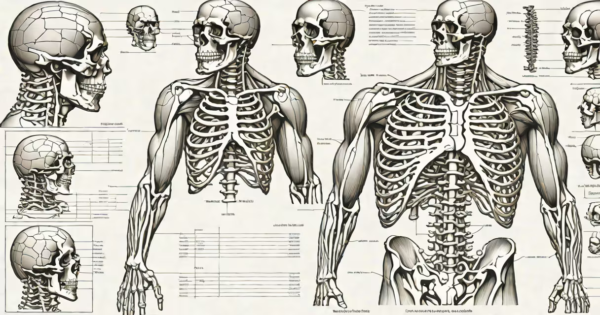
The human body torso is the central segment of the body that extends from the base of the neck to the pelvis, housing the heart, lungs, liver, stomach, intestines, and reproductive organs. It serves as the structural and functional core, anchoring the arms and legs while protecting important internal systems.
Located between the head and the lower limbs, the torso forms the trunk's foundation. It contains the thoracic cavity (chest) and abdominal cavity (belly), separated by the diaphragm-a dome-shaped muscle critical for breathing. The vertebral column runs through the center, providing stability and protection for the spinal cord. This region integrates movement, respiration, digestion, and circulation.
According to Gray's Anatomy, the torso accounts for approximately 40% of total body mass and supports over 60% of the body's musculature. Its integrity enables upright posture, balance during locomotion, and efficient force transfer during physical activity.
The torso consists of skeletal, muscular, nervous, and organ systems working in concert. The bony structure includes the sternum (breastbone), 12 pairs of ribs, and the vertebrae of the thoracic spine. These bones form a protective cage around the chest and upper abdomen.
The ribcage encloses the thoracic cavity. Each rib connects to the thoracic vertebrae posteriorly and to the sternum anteriorly via costal cartilage. The first seven pairs are true ribs; the next three are false ribs (attached indirectly); the last two are floating ribs (not attached to the sternum).
Internal organs include the heart and lungs in the thoracic cavity, and the liver, gallbladder, pancreas, stomach, small and large intestines, spleen, and kidneys in the abdominal cavity. The diaphragm separates these two regions and contracts during inhalation to draw air into the lungs.
Several distinct zones define the torso's organization:
| Region | Primary Structures | Functions |
|---|---|---|
| Thorax | Ribs, sternum, thoracic vertebrae, heart, lungs | Protects important organs, enables respiration, supports upper body |
| Abdomen | Abdominal wall muscles, liver, stomach, intestines, kidneys | Digestion, nutrient absorption, waste processing |
| Pelvis | Hip bones, sacrum, coccyx, reproductive organs | Supports weight, protects reproductive system, enables childbirth |
Each zone contributes uniquely to homeostasis. For example, the abdominal wall is composed of four layers: skin, subcutaneous tissue, superficial fascia, and deep fascia-each playing a role in structural support and sensory feedback.
During a routine physical exam, clinicians assess the torso's symmetry, respiratory rate, and abdominal tenderness. A palpable mass in the left upper quadrant may indicate splenic enlargement, while dullness on percussion over the right lower chest could signal pleural effusion.
Many assume the torso ends at the waistline. In reality, the anatomical boundary lies at the inferior border of the pelvic inlet-the point where the lumbar vertebrae meet the sacrum. The pelvis is part of the torso, not a separate entity.
Another error is equating the "core" with just the abdominal muscles. While rectus abdominis and transverse abdominis contribute, the core includes the erector spinae, multifidus, gluteals, and diaphragm. According to the American Council on Exercise, effective core training must engage all these muscles simultaneously.
A lesser-known fact: the human torso contains over 100 individual muscles. Among them, the quadratus lumborum stabilizes the lower back during standing and walking. Its dysfunction is linked to chronic low back pain, affecting up to 80% of adults at some point in life.
In surgical procedures, precise knowledge of the torso's anatomy prevents complications. During a laparoscopic cholecystectomy (gallbladder removal), surgeons enter through the umbilicus and go through using landmarks such as the falciform ligament and portal vein. Mistakes in identifying these structures can lead to bile duct injury, occurring in 0.5% to 1% of cases.
Consider a patient presenting with chest pain. A clinician evaluates the torso's vascular and neurological systems. If pain radiates to the left arm and jaw, it may indicate myocardial ischemia. The ECG shows ST-segment elevation, confirming acute coronary syndrome. Immediate intervention is required to prevent irreversible damage.
I once observed a cadaver dissection where a student misidentified the internal oblique muscle as the external oblique due to incorrect layer orientation. This mistake highlighted how subtle differences in muscle fiber direction affect function and diagnosis. Proper labeling ensures accurate learning and clinical application.
The ribs on a human body provide both protection and flexibility. They allow expansion during inhalation and recoil during exhalation. Fractures in the middle ribs are common in blunt trauma and may require imaging to rule out underlying lung injury.
Every action-from breathing to lifting weights-relies on coordinated function across the torso. Its design balances protection, mobility, and physiological efficiency. Without this integration, survival would be impossible.