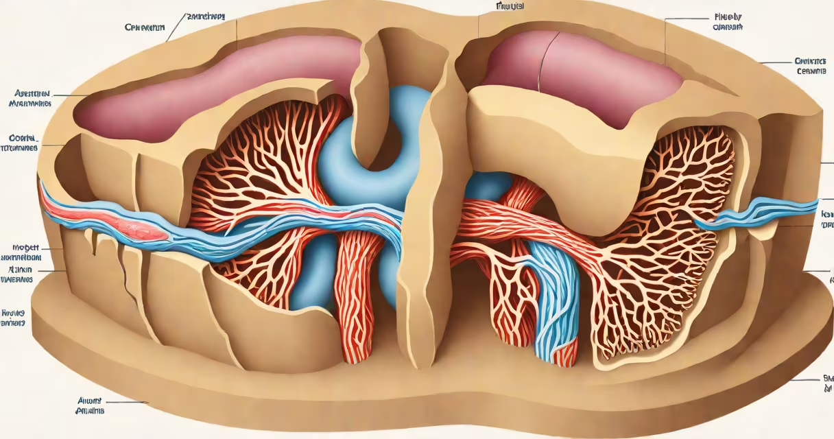
The organs in your abdominal cavity remain in position through a combination of muscular walls, fibrous connective tissues, and specialized membranes like the peritoneum. These structures form a changing system that stabilizes organs during movement, digestion, and changes in body posture.
Internal organ placement relies on both passive structural support and active muscular control. The abdominal wall-formed by layers of muscle and fascia-provides external containment. Inside, the peritoneum lines the abdominal cavity and anchors organs via ligaments and mesenteries. Connective tissue networks, including visceral fascia, further secure organs to their proper locations.
Without this integrated support system, organs would shift during daily activities. For example, the small intestine would move unpredictably during walking, disrupting nutrient absorption. The liver, spleen, and kidneys each rely on specific attachments to stay within their anatomical zones.
The primary mechanism holding organs in place is the peritoneum-a double-layered serous membrane lining the abdominal cavity and covering most abdominal organs. The parietal peritoneum adheres to the inner abdominal wall, while the visceral peritoneum envelops the organs themselves. Between these layers lies a thin fluid-filled space that reduces friction during organ movement.
Organs are tethered to the abdominal wall and each other through folds of peritoneum called mesentery, mesocolon, and omentum. These structures act like suspenders for internal organs. The transverse mesocolon, for instance, connects the transverse colon to the posterior abdominal wall and prevents it from sagging into the lower abdomen.
The abdominal muscles-rectus abdominis, external and internal obliques, and transversus abdominis-contract to increase intra-abdominal pressure. This pressure pushes organs upward and inward, helping maintain their alignment. During coughing or lifting, this action becomes especially critical to prevent organ prolapse.
Each layer of the abdominal support system plays a distinct role. The following table outlines the major elements involved:
| Structure | Function | Example Location |
|---|---|---|
| Peritoneum | Lines cavity and attaches organs via mesenteries | Wraps around intestines, liver, and stomach |
| Visceral fascia | Connective tissue network supporting organ surfaces | Surrounds kidneys and adrenal glands |
| Abdominal wall muscles | Provide compressive force and stability | Rectus abdominis and transversus abdominis |
| Retroperitoneal space | Contains organs anchored behind the peritoneum | Kidneys, pancreas, and aorta |
Some organs-like the kidneys and duodenum-are retroperitoneal, meaning they lie behind the peritoneal lining. These organs are secured by connective tissue rather than mesenteries. Their stability depends on direct attachment to the posterior abdominal wall, which prevents shifting during physical activity.
A common misconception is that organs float freely in the abdomen. They do not. Even though the peritoneal fluid allows limited gliding motion, organs remain firmly anchored. A study published in *Anatomical Record* confirms that the average human colon maintains its position within 1.2 cm of its baseline location during normal movement, thanks to mesenteric fixation points.
Many assume that the abdominal muscles alone hold organs in place. While they contribute, they are not the primary stabilizers. The peritoneum and fascial layers bear the majority of the mechanical load. Without these, even strong abdominal muscles couldn't prevent organ displacement during sudden movements.
Another error is equating organ support with ligament strength. While ligaments like the gastrohepatic ligament stabilize parts of the liver, they don't provide organ positioning. Instead, the entire system functions as a network. If one component fails-such as a torn mesentery-other structures compensate, but severe trauma can lead to organ misplacement.
One non-obvious fact: the peritoneum is not just a passive sheet. It actively secretes lubricating fluid and contains immune cells. When inflamed due to infection or injury, it can cause adhesions-bands of scar tissue that bind organs together. These adhesions may restrict normal organ movement and cause chronic pain, demonstrating how important the peritoneum's integrity is.
A hernia occurs when weakened abdominal wall tissue allows organs to protrude. For example, an inguinal hernia results when part of the intestine pushes through a weak spot in the lower abdominal wall. This happens because the muscular and fascial layers have degraded over time or under strain.
Consider a 55-year-old man who lifts heavy objects regularly. His transversus abdominis muscle has atrophied slightly due to aging. Over time, the connective tissue supporting the spermatic cord weakens. During a lift, increased intra-abdominal pressure forces the small intestine through the internal ring into the scrotum. This is a direct failure of the support system-specifically, the combination of muscle weakness and fascial degeneration.
Repair involves reinforcing the abdominal wall with synthetic mesh. The procedure restores the mechanical barrier that normally prevents organ displacement. Without surgical intervention, the hernia can grow larger and lead to bowel obstruction.
Organ displacement is rare in healthy individuals but can occur due to trauma, surgery, or congenital defects. For instance, malrotation of the intestines during fetal development can result in inadequate mesenteric attachment. This condition increases the risk of volvulus-twisting of the bowel-which cuts off blood flow and requires emergency surgery.
Post-surgical patients often experience temporary organ instability. After laparoscopic procedures, residual gas in the peritoneal cavity causes bloating and altered organ positioning. This is why patients are advised to avoid strenuous activity for several weeks-allowing connective tissues and muscles to re-establish tension.
I once observed a patient with a history of multiple abdominal surgeries develop recurrent adhesions. Despite normal muscle strength, his small intestine was bound to the anterior abdominal wall by dense scar tissue. He experienced intermittent bowel obstruction. Laparoscopic lysis of adhesions restored normal function. This case illustrates that even intact muscles cannot overcome compromised connective tissue support.
Understanding what holds your organs in place reveals the complexity of internal anatomy. It's not one structure but a coordinated system of membranes, muscles, and connective tissues working in tandem. Maintaining this system through healthy weight, core strengthening, and avoiding repetitive strain helps preserve long-term organ stability.