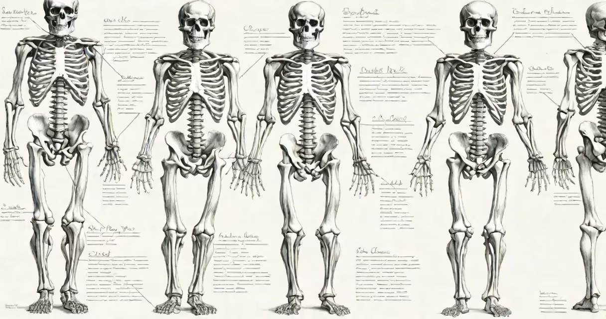
Human bones and muscles form the body's structural and locomotive structure, enabling posture, movement, and protection of internal organs. Together, they make up the musculoskeletal system, which allows humans to stand, walk, lift, and perform complex physical tasks.
The skeletal system consists of 206 bones in the adult human body, arranged into axial (skull, spine, ribcage) and appendicular (limbs, shoulder girdle, pelvic girdle) divisions. These bones provide rigidity, store minerals like calcium and phosphorus, and produce blood cells in the bone marrow. The femur, the longest bone at approximately 48 cm in average adults, supports nearly all body weight during standing and walking.
The muscular system comprises over 600 muscles, primarily skeletal muscles attached to bones via tendons. These muscles contract using actin and myosin filaments, generating force through biochemical energy from ATP. For example, the biceps brachii contracts to flex the forearm at the elbow, allowing lifting actions. Skeletal muscles account for about 40% of total body mass in healthy adults.
Bones serve as levers, while muscles act as motors. When a muscle contracts, it pulls on a tendon, which attaches to a bone. This creates movement around a joint, such as the knee bending when the quadriceps contract and the hamstrings relax. This coordinated action is controlled by the nervous system, which sends signals through motor neurons to initiate contraction.
A single movement often involves multiple muscle groups. Flexing the arm requires the biceps to contract while the triceps relaxes. This antagonistic pair ensures smooth motion and prevents joint damage. Without this balance, movements would be jerky or unstable.
For example, during a push-up, the pectoralis major and anterior deltoid contract to extend the shoulder, the triceps extend the elbow, and the rectus abdominis stabilizes the trunk. Simultaneously, the spine remains aligned due to the support of the vertebral column and surrounding paraspinal muscles.
There are three main types of bones: long bones (e.g., femur), flat bones (e.g., sternum), and irregular bones (e.g., vertebrae). Long bones have a shaft (diaphysis) and two ends (epiphyses), with spongy bone tissue inside and compact bone on the surface. The epiphyseal plate, a layer of cartilage near the ends, allows growth in childhood and closes during puberty.
There are three types of muscle fibers: Type I (slow oxidative), Type IIa (fast oxidative), and Type IIb (fast glycolytic). Type I fibers are fatigue-resistant and used in endurance activities like long-distance running. Type IIb fibers generate high force quickly but fatigue rapidly, ideal for sprinting or heavy lifting.
| Type | Function | Example | Energy Source |
|---|---|---|---|
| Skeletal | Voluntary movement, posture | Biceps brachii | ATP, glucose, fatty acids |
| Smooth | Involuntary functions (digestion, blood flow) | Intestinal wall | Glucose, oxygen |
| Cardiac | Heart contractions | Myocardium | Fatty acids, glucose |
While most people think of muscles as visible and voluntary, smooth and cardiac muscles operate automatically. Smooth muscle lines the digestive tract and blood vessels, regulating flow without conscious control. Cardiac muscle forms the heart wall and maintains rhythmic contractions throughout life.
Many assume bones are inert and static. In reality, bones are living tissues that remodel continuously. Osteoblasts build new bone, while osteoclasts break down old bone. This process, called bone remodeling, occurs every 10 years across the entire skeleton and is critical for healing fractures and maintaining mineral balance.
Another misconception is that muscles grow larger simply by lifting weights. Muscle hypertrophy results from microtears in muscle fibers during resistance training, followed by repair and protein synthesis. A study published in the Journal of Applied Physiology found that consistent resistance training increases muscle fiber diameter by 10-15% over 12 weeks in trained individuals.
People often believe that stretching before exercise prevents injury. Research from the American College of Sports Medicine shows that changing stretching (movement-based) improves performance and reduces injury risk more effectively than static stretching done before activity. Static stretching is better suited for post-exercise recovery.
I once observed a patient recovering from a tibial fracture who had undergone surgical fixation with a titanium rod. Over six months, imaging showed progressive bone callus formation around the implant, confirming active remodeling. The patient regained full mobility after physical therapy focused on muscle reactivation and joint stability.
Understanding human bones and muscles is essential in fields like orthopedic surgery, sports medicine, and rehabilitation. For instance, athletes train specific muscle groups to improve performance. A sprinter develops explosive power in the gluteus maximus and quadriceps, while a pianist trains fine motor control in the hand muscles.
Human bones and muscles are not passive structures. They adapt to load, respond to injury, and evolve with training. Their interaction underpins nearly every physical act-from blinking to running a marathon. Recognizing their roles and limitations enhances health, performance, and injury prevention.