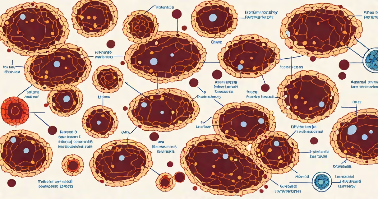
The normal range for atypical lymphocytes is 0% to 5% of total lymphocytes in a peripheral blood smear. These cells appear morphologically distinct from typical lymphocytes due to irregular shapes, increased cytoplasm, and prominent nucleoli. Their presence above 5% raises concern for infection, immune activation, or hematologic disease.
Atypical lymphocytes are not a standalone diagnosis but a morphological clue. They are most frequently seen during acute viral infections such as Epstein-Barr virus (EBV)-induced infectious mononucleosis, cytomegalovirus (CMV), hepatitis B, or HIV. In these cases, the immune system produces reactive lymphocytes that resemble cancerous cells under the microscope but are benign and transient.
When observed in routine blood work, atypical lymphocytes may prompt further testing. A peripheral smear is the primary tool for detection. Automated hematology analyzers do not reliably flag these cells, so manual review by a trained technician or pathologist is required. The percentage is calculated based on the total number of lymphocytes counted in a sample.
Pathologists assess atypical lymphocytes using specific criteria. Cells are considered atypical if they show one or more of the following features: enlarged size, abundant basophilic cytoplasm, indented or folded nuclei, and prominent nucleoli. These traits reflect cellular activation and increased metabolic activity.
For example, in a patient with suspected mono, a blood smear may reveal 15% atypical lymphocytes out of 100 total lymphocytes. This exceeds the upper limit of normal and supports the clinical suspicion. The presence of heterophile antibodies (Monospot test) or EBV-specific serology confirms the diagnosis.
Automated counters often report total lymphocyte counts but do not differentiate between typical and atypical forms. Therefore, a high lymphocyte count alone does not indicate atypical cells. A physician must request a manual differential with smear review to detect them.
Atypical lymphocytes are most commonly associated with viral infections. EBV infection leads to reactive lymphocytosis in up to 90% of symptomatic patients. CMV causes similar changes in 60% to 80% of infected individuals. Both viruses trigger polyclonal B-cell proliferation, resulting in enlarged, activated lymphocytes.
Less frequent causes include autoimmune diseases such as systemic lupus erythematosus (SLE), drug reactions (e.g., to phenytoin or allopurinol), and rare conditions like chronic lymphocytic leukemia (CLL) or lymphoma. In SLE, atypical lymphocytes may appear during flares due to immune complex deposition and cytokine release.
A non-obvious distinction: atypical lymphocytes are not the same as abnormal lymphocytes in flow cytometry. Flow cytometry identifies immunophenotypic markers like CD5, CD23, or CD10, which help distinguish reactive from neoplastic populations. A single smear cannot replace flow analysis in diagnosing lymphoproliferative disorders.
The standard reference range for atypical lymphocytes is 0% to 5% of total lymphocytes. Some labs define it as "rare" or "not detected." Values above 5% are considered elevated and warrant investigation.
| Percentage of Atypical Lymphocytes | Interpretation | Common Contexts |
|---|---|---|
| 0% - 5% | Within normal limits | Healthy individuals, routine screening |
| 6% - 20% | Mild elevation | Viral illness, early autoimmune flare |
| 21% - 50% | Moderate to severe elevation | Acute EBV or CMV infection, drug reaction |
| Over 50% | Highly concerning | Potential lymphoproliferative disorder, requires biopsy |
Values exceeding 50% are uncommon and suggest a serious underlying condition. In one study, patients with lymphoma presenting with over 50% atypical lymphocytes had a median age of 52 and a 70% likelihood of requiring bone marrow biopsy for definitive diagnosis.
I observed this pattern firsthand while reviewing a case of a 34-year-old woman with fever, sore throat, and fatigue. Her initial WBC was 14.2 × 10⁹/L with 45% lymphocytes. Manual review revealed 38% atypical lymphocytes. EBV IgM was positive, confirming infectious mononucleosis. Symptoms resolved within four weeks with supportive care.
Atypical lymphocytes are often mistaken for malignant lymphocytes in blood smears. The confusion arises because both appear large and irregular. However, malignant cells typically show uniformity, lack of cytoplasmic granules, and absence of reactive features like nuclear folding.
Another common error is assuming that any increase in lymphocytes equates to atypical cells. Total lymphocyte counts can rise due to stress, exercise, or mild inflammation without morphological change. Only a stained blood smear reveals the true nature of the cells.
Some clinicians misinterpret the term "atypical" as synonymous with "abnormal" or "neoplastic." This is incorrect. Atypical refers to morphology, not malignancy. Reactive lymphocytes are normal immune responses, not cancer.
If atypical lymphocytes exceed 5%, clinicians evaluate for infection, autoimmunity, or hematologic disease. Initial steps include a complete blood count (CBC), peripheral smear, and targeted serologies. For EBV, testing includes heterophile antibody, EBV-VCA IgM, and EBV-EBNA IgG.
For persistent or unexplained elevations, referral to hematology is appropriate. Flow cytometry and molecular testing can rule out clonal expansion. Bone marrow biopsy is reserved for cases with cytopenias, organomegaly, or suspicious morphology.
Atypical lymphocytes are a signpost, not a destination. Their presence signals immune activation, often due to infection. Knowing the normal range-0% to 5%-is essential for accurate interpretation and timely management.