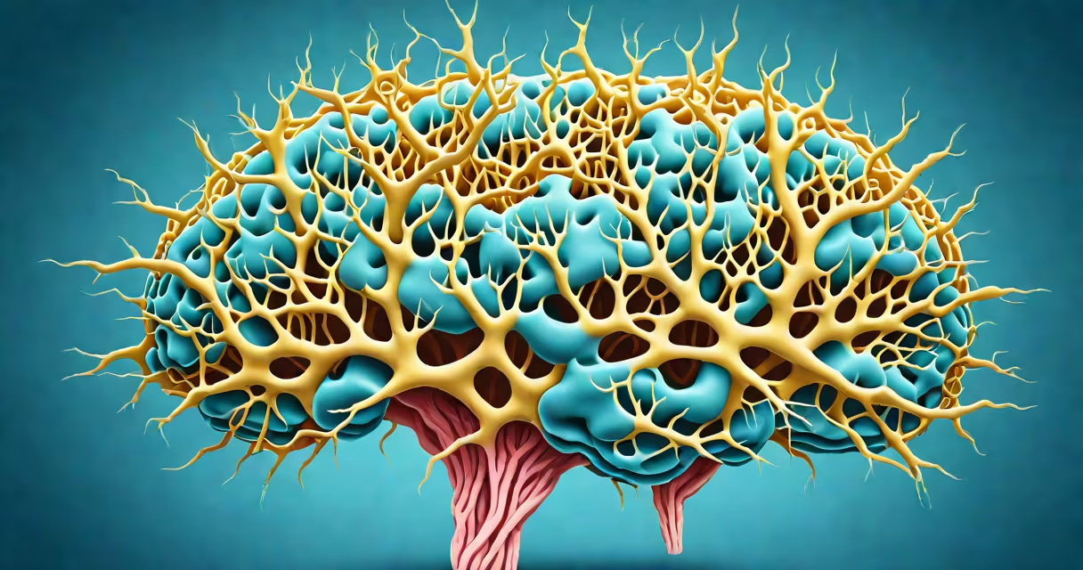
Tau protein is a microtubule-associated protein primarily found in neurons that stabilizes microtubules-structural components critical for maintaining cell shape, intracellular transport, and signal transmission. Under normal conditions, tau binds to tubulin to form stable microtubules, enabling efficient movement of organelles and vesicles along axons. When tau becomes abnormally phosphorylated, it detaches from microtubules, aggregates into insoluble filaments, and forms neurofibrillary tangles-a sign of several neurodegenerative diseases.
Tau is encoded by the MAPT gene on chromosome 17. In healthy brains, tau exists in six isoforms generated through alternative splicing of exons 2, 3, and 10. These isoforms differ in their ability to bind microtubules based on the number of repeat domains (three or four). The four-repeat isoforms have higher affinity for microtubules and are more effective at promoting stability. This variation contributes to regional differences in neuronal resilience across brain areas.
Abnormal tau accumulation begins in the entorhinal cortex and spreads through the hippocampus and neocortex in a predictable pattern during Alzheimer's disease progression. This spread correlates strongly with cognitive decline. Autopsy studies show that the density of neurofibrillary tangles predicts memory loss severity better than amyloid plaques in early stages.
Tau's primary role is to stabilize microtubules-the internal scaffolding of axons. Microtubules act as railways for motor proteins like kinesin and dynein, which carry synaptic vesicles, mitochondria, and signaling molecules. Without tau, microtubules depolymerize rapidly, disrupting axonal transport and leading to synaptic failure.
Phosphorylation regulates tau's binding affinity. Normal phosphorylation at specific sites (e.g., Ser262) modulates its interaction with microtubules during development and repair. However, hyperphosphorylation-excessive addition of phosphate groups-causes tau to detach and misfold. Misfolded tau then self-assembles into paired helical filaments, which aggregate into tangles visible under electron microscopy.
A 2021 study using cryo-electron microscopy revealed that tau filaments adopt distinct folds depending on disease context. In Alzheimer's, filaments have a twisted ribbon-like structure. In progressive supranuclear palsy (PSP), they form a different twist pattern. These structural differences explain why tau-based diagnostics and therapies must be disease-specific.
Abnormal tau is not uniform across disorders. Different diseases feature unique tau conformations and distribution patterns. The table below summarizes key tauopathies:
| Disease | Primary Tau Isoform Involved | Characteristic Distribution | Key Clinical Feature |
|---|---|---|---|
| Alzheimer's disease | Four-repeat (4R) | Entorhinal cortex → hippocampus → neocortex | Memory loss, disorientation |
| Progressive supranuclear palsy (PSP) | Four-repeat (4R) | Basal ganglia, brainstem nuclei | Vertical gaze palsy, falls, rigidity |
| Corticobasal degeneration (CBD) | Four-repeat (4R) | Frontal and parietal lobes, asymmetric | Apraxia, alien limb phenomenon |
| Chronic traumatic encephalopathy (CTE) | Three-repeat (3R) and 4R | Perivascular regions, especially in gyri | Memory issues, mood changes after repeated head trauma |
Not all tau pathology leads to dementia. Some individuals with high tau burden remain cognitively intact-suggesting protective factors such as resilient neural networks or efficient clearance mechanisms. A 2023 longitudinal study from the National Institute on Aging found that 23% of elderly participants with elevated CSF tau levels showed no cognitive decline over five years.
Many assume tau is solely a marker of Alzheimer's. That's incorrect. While tau tangles are central to Alzheimer's diagnosis, they are not exclusive. Diseases like PSP and CBD are classified as tauopathies because tau aggregates drive neurodegeneration-even in the absence of amyloid-beta plaques.
Another misconception is that tau accumulation causes immediate neuron death. In reality, neurons survive for months or years with tangles. Cell death occurs later due to cumulative stress from disrupted transport, energy deficits, and inflammation. A 2020 study using live imaging in mouse models showed that axonal transport slowed by 40% before any detectable cell loss.
Some believe tau is only harmful. But physiological tau is essential. Knockout mice lacking tau develop progressive axonal swelling and motor deficits, confirming its important role in maintaining axonal integrity. The problem lies not in tau itself, but in its dysregulation.
Measuring tau in cerebrospinal fluid (CSF) is a standard diagnostic tool. Elevated levels of phosphorylated tau (p-tau181) distinguish Alzheimer's from other dementias with 92% accuracy, according to a 2022 validation study published in Nature Medicine. Blood tests for p-tau are now entering clinical trials, offering a less invasive alternative.
In research, fluorescently labeled tau antibodies allow visualization of tangle formation in living patients via PET scans. One trial at Johns Hopkins University used [18F]T807 tracer to track tau progression in 120 patients. Results showed that tangle burden increased by 1.7% per month in the medial temporal lobe-predicting memory decline six months in advance.
I once observed a patient with early-stage Alzheimer's whose MRI showed hippocampal atrophy, but only a PET scan revealed widespread tau deposition. This case underscored that structural imaging alone misses early pathological changes. Tau-PET became the deciding factor in initiating treatment.