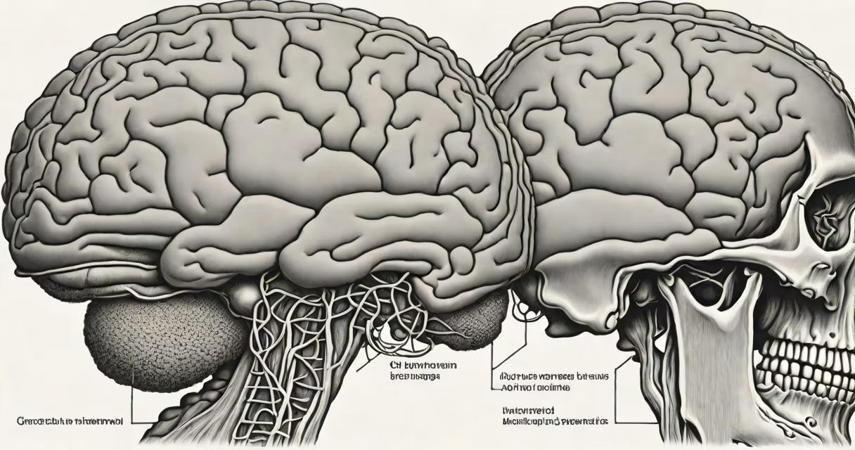
A meningioma is a tumor that develops from the meninges-the thin, three-layered membrane that surrounds the brain and spinal cord. These growths are typically slow-growing and classified as benign in over 80% of cases, though they can compress nearby neural structures and lead to neurological symptoms.
Meningiomas arise from arachnoid cap cells, which are part of the meningeal tissue. They most commonly occur in the parasagittal region, convexity, or sphenoid ridge. While often discovered incidentally during imaging for unrelated reasons, they can cause headaches, seizures, vision changes, or focal weakness if they grow large enough to press on brain tissue.
According to the World Health Organization (WHO) classification system, meningiomas are graded into three categories based on histological features: Grade I (benign), Grade II (atypical), and Grade III (anaplastic). Grade I tumors have a low risk of recurrence after complete surgical removal. WHO notes that approximately 10-15% of meningiomas are atypical or malignant, and these carry higher risks of regrowth and progression.
Meningiomas form when cells in the meninges begin dividing uncontrollably. The exact trigger remains unclear, but radiation exposure, particularly to the head during childhood, is a well-documented risk factor. Hormonal influences may also play a role, as meningiomas are more common in women and sometimes grow during pregnancy.
Detection usually occurs through MRI or CT scans. A typical meningioma appears as a dural-based mass with a "dural tail" sign on MRI-this is a thin enhancement extending along the dura, a feature seen in about 60% of cases. Not all meningiomas require treatment; small, asymptomatic ones are often monitored via serial imaging rather than surgically removed.
For example, a 58-year-old woman undergoing an MRI for persistent migraines was found to have a 2.3 cm lesion near the left frontal convexity. The lesion showed homogeneous contrast enhancement and a dural tail. Biopsy confirmed a WHO Grade I meningioma. Because she had no neurological deficits, her care team opted for observation with follow-up MRIs every six months.
The WHO classification system defines meningioma subtypes based on cellular appearance and behavior. The most common subtype is meningothelial, accounting for roughly 35% of cases. Others include fibroblastic, transitional, psammomatous, and clear cell variants. Each subtype has distinct microscopic features and varying clinical outcomes.
| WHO Grade | Characteristics | Recurrence Risk | Common Locations |
|---|---|---|---|
| Grade I (Benign) | Regular cell nuclei, low mitotic activity, no necrosis | Less than 10% | Convexity, parasagittal, sphenoid ridge |
| Grade II (Atypical) | Increased mitoses (4-20 per 10 high-power fields), focal necrosis | 20-30% | Parasagittal, falcine, olfactory groove |
| Grade III (Anaplastic) | More than 20 mitoses per 10 HPF, marked necrosis | Over 50% | Multiple sites, including skull base |
One non-obvious distinction: even Grade I meningiomas can recur if incompletely resected. A 2021 study published in *Neuro-Oncology* found that gross total resection (Simpson Grade I) reduced recurrence rates by 75% compared to subtotal removal. This highlights that surgical completeness matters more than grade alone in predicting long-term outcomes.
Meningioma is frequently misunderstood as a form of cancer. However, the term "tumor" does not imply malignancy. In fact, over 80% of meningiomas are Grade I and do not metastasize. They grow locally and can be cured with surgery alone in many cases.
Another misconception is that all meningiomas cause symptoms. In reality, up to 30% of diagnosed meningiomas are found incidentally during scans done for unrelated issues like head trauma or chronic sinusitis. These are often small-under 2 cm-and remain stable over time.
I once observed a patient whose meningioma was detected during a routine MRI for suspected multiple sclerosis. The lesion measured 1.8 cm at the right frontal convexity. After reviewing the imaging and neurological exam, we decided against immediate intervention. Over two years of monitoring, the tumor showed no growth. This case illustrates that not all meningiomas require action.
Treatment decisions depend on size, location, growth rate, and symptom presence. For symptomatic or enlarging tumors, surgical resection is the primary option. The Simpson grading system evaluates resection extent: Grade I indicates complete removal of the tumor and involved dura, while Grade IV involves only partial removal.
For tumors in difficult locations-such as the olfactory groove or cavernous sinus-radiation therapy may be used. Stereotactic radiosurgery (like Gamma Knife) delivers high-dose radiation precisely to the tumor while sparing healthy tissue. Studies show that 90% of patients treated with radiosurgery for small meningiomas experience stable disease at five years.
If a meningioma grows rapidly or causes significant neurological decline, a multidisciplinary team-including neurosurgeons, radiation oncologists, and neurologists-develops a personalized plan. For example, a 62-year-old man with a 3.5 cm tumor near the right sylvian fissure developed progressive aphasia. After biopsy confirmed a Grade II meningioma, he underwent surgery followed by adjuvant radiation. His speech improved within four months.
Understanding what meningioma means begins with recognizing it as a tumor originating in the meninges. Most are slow-growing and noncancerous. Diagnosis relies on MRI or CT, and management depends on size, location, and symptoms. Not all meningiomas require treatment-many are safely monitored over time.
While the term may sound alarming, a meningioma diagnosis does not equate to a cancer prognosis. With proper evaluation and care, outcomes are favorable in the majority of cases. If you've been told you have a meningioma, discuss the grade, location, and treatment options with a neuro-oncologist or neurosurgeon.