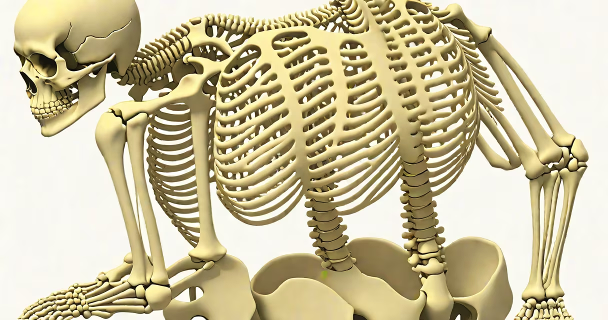
The back of a skeleton refers to the posterior aspect of the human bony structure, dominated by the vertebral column, thoracic cage, and scapulae. It provides structural support, protects the spinal cord, and enables upright posture and complex motion.
This region includes 33 individual vertebrae arranged into five regions: cervical (7), thoracic (12), lumbar (5), sacral (5 fused), and coccygeal (4 fused). These bones form the spine, which runs from the base of the skull to the tailbone. The thoracic vertebrae connect to 12 pairs of ribs, forming the ribcage that encloses important organs like the heart and lungs.
The scapulae, or shoulder blades, sit flat against the upper back, anchoring muscles involved in arm movement. They articulate with the humerus at the glenohumeral joint and provide attachment points for over 20 muscles. Together, these elements create a rigid yet flexible scaffold essential for locomotion and stability.
Movement at the back of the skeleton occurs through controlled articulation between vertebrae. Intervertebral discs act as shock absorbers between adjacent vertebrae, allowing limited flexion, extension, lateral bending, and rotation. The lumbar spine bears most of the body's weight during standing and walking.
Nerve roots exit the spinal cord through intervertebral foramina between each pair of vertebrae. Damage to these pathways-such as from herniated discs or spinal stenosis-can cause pain, numbness, or weakness in the limbs. The spinal cord itself extends from the brainstem to the level of L1, protected within the vertebral canal.
Postural control relies heavily on the back's musculature. Deep stabilizing muscles like the multifidus and transverse abdominis work with larger extensors such as the erector spinae to maintain alignment. Without this system, balance would fail under normal daily loads.
The following table outlines major structures visible in the back of a skeleton and their functions:
| Structure | Number of Elements | Primary Function |
|---|---|---|
| Vertebral column | 33 bones (5 regions) | Supports body weight, houses spinal cord, enables movement |
| Ribcage | 12 pairs | Protects heart and lungs; assists in breathing mechanics |
| Scapulae | 2 bones | Anchor upper limb muscles; allow wide range of arm motion |
| Sacrum | td>1 bone (fused)Forms posterior wall of pelvis; connects spine to hip bones |
Each component contributes to both protection and mobility. For example, the thoracic vertebrae have facets that lock onto rib heads, creating a stable yet slightly mobile cage. This design allows for respiratory expansion while resisting deformation under load.
Many assume the back of a skeleton is merely a stack of bones. In reality, it integrates changing systems: ligaments stabilize joints, tendons transmit muscle force, and nerves regulate sensation and motor control. The spine is not a rigid rod-it bends, twists, and adapts to terrain and activity.
A frequent error is equating the spine with the entire back. While the vertebral column is central, the back also includes soft tissue attachments visible only in living bodies. In anatomical models, the absence of skin, fat, and muscle makes the skeleton appear simpler than it is in life.
Another confusion arises between the back of a skeleton and the back of a cadaver. A preserved cadaver shows muscle layers, blood vessels, and nerve trunks. In contrast, a cleaned skeleton displays only bone surfaces, making it easier to study articulations but harder to grasp biomechanical function.
In 2021, a 45-year-old office worker presented with chronic lower back pain. Imaging revealed degenerative disc disease at L4-L5. The patient's symptoms included radiating pain down the left leg and diminished ankle reflexes-classic signs of sciatic nerve compression.
Anatomical analysis confirmed that the L4-L5 intervertebral disc had narrowed, reducing space in the neural foramen. This narrowed passage compressed the L5 nerve root, leading to sensory loss and muscle weakness. The back of the skeleton, particularly the lumbar vertebrae and exiting nerve roots, was critical in diagnosing the issue.
After conservative treatment failed, surgery was performed to decompress the nerve. Post-op imaging showed restored foraminal width and relief of pressure. The patient regained strength and returned to work within six months.
This case illustrates how understanding the back of a skeleton informs medical decisions. Radiologists and surgeons rely on precise knowledge of bony landmarks to plan interventions safely.
The back of a skeleton connects closely with other regions. The cervical spine continues upward from the skull, forming the atlanto-occipital joint that allows nodding motion. The thoracic spine curves slightly forward (kyphosis), balancing the lordotic curve of the lumbar spine.
One non-obvious fact: the sacrum is not a single bone but a fusion of five vertebrae. This fusion begins around age 16 and completes by age 25. The resulting structure resists torsional forces during walking and running, distributing weight evenly across the pelvis.
During my time working in a university anatomy lab, I observed a dissection where students removed the skin and muscles from a cadaver's back. The underlying bone pattern-especially the spinous processes and transverse processes-revealed how muscle attachment sites evolved for bipedalism. These features are absent in quadrupedal animals, underscoring human-specific adaptations.
Understanding the back of a skeleton is foundational for medicine, physical therapy, and biomechanics. It is not just a passive frame but an active system shaped by evolution, development, and daily use.