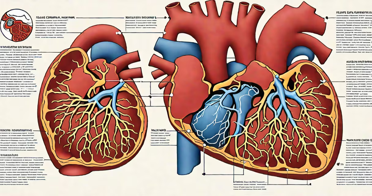
Left sided heart failure causes fluid to accumulate in the lungs due to impaired pumping of the left ventricle, resulting in pulmonary congestion, shortness of breath, and abnormal lung sounds. This occurs when the left ventricle cannot eject blood effectively during systole, increasing pressure in the pulmonary circulation.
The primary mechanism involves reduced cardiac output and elevated left ventricular end-diastolic pressure. As pressure rises in the left atrium and pulmonary capillaries, fluid leaks into the alveoli and interstitial spaces. This process is driven by Starling forces-when hydrostatic pressure exceeds oncotic pressure, fluid moves out of capillaries into surrounding tissues.
According to the American Heart Association, left sided heart failure accounts for approximately 50% of all heart failure cases. It often stems from coronary artery disease, hypertension, or myocardial infarction. The condition progresses through stages defined by the New York Heart Association (NYHA) functional classification, with symptoms worsening from class I (no limitation) to class IV (symptoms at rest).
When the left ventricle fails, it cannot contract forcefully enough to push blood forward into the systemic circulation. Blood backs up into the left atrium, then into the pulmonary veins and capillaries. Elevated pulmonary capillary pressure leads to transudation of fluid into lung tissue.
This fluid accumulation impairs gas exchange. Oxygen diffusion across the alveolar-capillary membrane slows, reducing arterial oxygen saturation. Patients experience exertional dyspnea-shortness of breath during activity-and later, dyspnea at rest. They may also develop orthopnea, needing multiple pillows to sleep comfortably.
A non-obvious distinction: left sided heart failure does not initially cause peripheral edema. That typically appears in right sided failure. Confusing the two delays diagnosis. A patient with leg swelling and ascites likely has right-sided or biventricular failure, not isolated left-sided dysfunction.
Shortness of breath is the most common symptom. It begins with exertion and progresses to occur at rest. Patients describe it as "air hunger" or feeling "like something's crushing my chest."
Clinicians listen for crackles-fine, discontinuous popping sounds heard on auscultation, especially at the lung bases. These indicate fluid in the alveoli. A third heart sound (S3 gallop) may be audible during diastole, signaling rapid ventricular filling under high pressure.
Patients often report waking at night gasping for air-paroxysmal nocturnal dyspnea. This occurs because lying flat increases venous return to the heart, worsening pulmonary congestion. Sitting upright reduces this effect by lowering central venous pressure.
| Symptom | Underlying Mechanism | Typical Onset |
|---|---|---|
| Dyspnea on exertion | Reduced cardiac output limits oxygen delivery | Early stage |
| Orthopnea | Increased venous return in supine position | Progressive |
| Paroxysmal nocturnal dyspnea | Fluid redistribution during sleep | Advanced |
| Crackles on exam | Pulmonary interstitial edema | Present in 60-70% of cases |
Many assume that all heart failure presents with fatigue and swelling. But left sided failure primarily affects the lungs, not the extremities. Peripheral edema suggests right-sided involvement or volume overload.
Another error: equating left sided heart failure with angina. Angina is chest pain from myocardial ischemia due to inadequate coronary perfusion. While both conditions can coexist-especially in patients with coronary artery disease-they are distinct. Angina causes pain, not pulmonary congestion.
I once saw a 68-year-old man with persistent cough and fatigue. He had normal blood pressure and no leg swelling. His ECG showed old inferior MI. A chest X-ray revealed bilateral pleural effusions and Kerley B lines-indicative of pulmonary edema. He was diagnosed with left sided heart failure, not pneumonia. Treating him for infection delayed recovery until the correct diagnosis was made.
Echocardiography is the gold standard for assessing left ventricular function. A reduced ejection fraction (<40%) confirms systolic heart failure. A preserved ejection fraction (>50%) points to diastolic failure, where the ventricle stiffens and fills poorly despite normal contraction.
B-type natriuretic peptide (BNP) levels help differentiate heart failure from other causes of dyspnea. Levels above 100 pg/mL suggest heart failure. A level below 100 makes it unlikely. BNP is released in response to stretch in the ventricular wall-directly reflecting volume overload.
For patients with suspected left sided failure, a chest X-ray shows cardiomegaly, increased vascular markings, and possibly pleural effusions. The "bat wing" appearance of pulmonary edema is a classic sign. In severe cases, the heart silhouette enlarges due to left ventricular dilation.
Treatment focuses on reducing preload and afterload. Diuretics like furosemide reduce fluid volume. ACE inhibitors or ARBs lower systemic vascular resistance. Beta-blockers such as carvedilol improve long-term survival by reducing heart rate and myocardial oxygen demand.
Without treatment, left sided heart failure worsens. Mortality rates exceed 50% at five years post-diagnosis, according to the Framingham Heart Study. Early intervention improves outcomes. Patients who adhere to medication regimens and lifestyle changes live longer and have fewer hospitalizations.