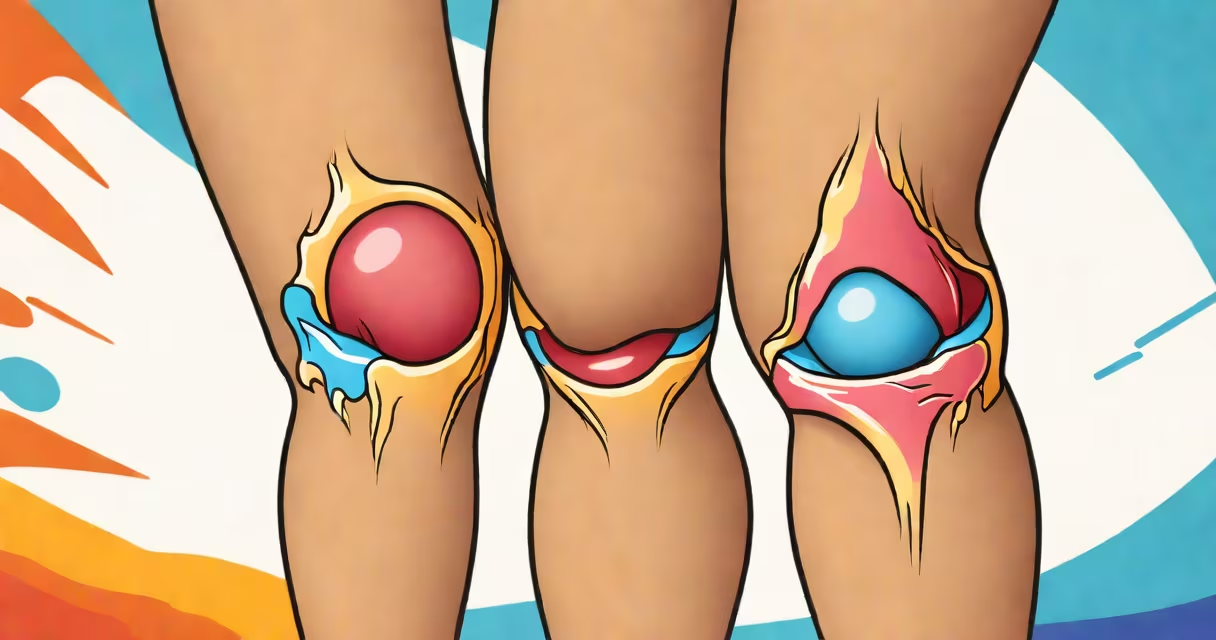
A torn medial collateral ligament (MCL) typically shows visible swelling, bruising, and tenderness along the inner side of the knee. The injury often results from a direct blow to the outer knee or sudden twisting during sports. Swelling may appear within hours, and bruising can spread down the lower leg over 24-48 hours.
The MCL stabilizes the inner knee joint. When damaged, it causes instability and pain when weight is applied. A grade 1 tear involves mild stretching, while a grade 3 tear means complete rupture. Most people describe a popping sensation at the time of injury, though not everyone hears or feels it.
Imaging confirms the extent. MRI scans show fluid buildup and disrupted ligament fibers. X-rays rule out bone fractures but don't visualize soft tissue. Ultrasound can detect tears in real time but requires a trained operator. An experienced orthopedic surgeon can diagnose a tear through physical exams, including valgus stress testing.
Swelling is the most immediate sign. It appears within 6-12 hours after injury and affects the entire knee joint. The area around the medial joint line-the inner edge of the knee-feels warm and tender to touch. Patients often report difficulty walking or bearing weight due to pain.
Bruising develops over 1-2 days. It starts near the inner knee and may extend down the shin. The color shifts from red-purple to blue-green to yellow as hemoglobin breaks down. This progression follows the path of gravity and tissue planes, not necessarily the injury site.
Visible deformity is rare unless the tear is severe. In grade 3 injuries, the knee may feel loose or give way during movement. Some patients report a "wobbly" sensation when standing or turning. These signs are more common in athletes who return to play too soon.
Doctors use the Lachman test and anterior drawer test to assess ligament stability. For MCL injuries, the valgus stress test is key. The examiner applies pressure to the outer knee while the leg is bent at 30 degrees. Pain or increased gapping on the inner side indicates MCL damage.
MRI scans provide detailed images of soft tissues. A torn MCL appears as a discontinuous signal in the ligament, with surrounding edema (fluid). According to the American Academy of Orthopaedic Surgeons (AAOS), MRI detects MCL tears with 95% accuracy when performed within 7 days of injury.
Differentiating MCL tears from ACL injuries is critical. ACL tears cause a "popping" sound and rapid swelling. They affect knee rotation and stability more severely. MCL tears are less likely to cause joint locking. Misdiagnosis leads to improper treatment-especially in athletes returning to sport.
| Type | Grade | Visual Signs | Recovery Time |
|---|---|---|---|
| MCL Tear | 1 | Mild swelling, localized tenderness | 2-4 weeks |
| MCL Tear | 2 | Significant swelling, bruising, limping | 4-6 weeks |
| MCL Tear | 3 | Severe swelling, joint instability, inability to bear weight | 8-12 weeks |
Many assume a visible bruise means a serious injury. But bruising can occur with minor sprains. Conversely, some grade 3 tears show little external change because blood pools beneath deep tissue layers. This delays diagnosis.
Another myth is that MCL tears always require surgery. The AAOS states that 80% of MCL injuries heal with conservative care-rest, bracing, and physical therapy. Surgery is reserved for cases with concurrent ACL damage or chronic instability.
I once evaluated a high school football player who claimed he "just twisted his knee." He had no bruising but reported inner knee pain when pivoting. On exam, valgus stress caused pain at 30 degrees of flexion. MRI confirmed a grade 2 MCL tear. He returned to play in six weeks with a hinged brace and strengthening exercises.
Most people recover full function within 6-12 weeks. Early rehabilitation focuses on reducing swelling and restoring range of motion. Ice, compression, and elevation reduce inflammation. Weight-bearing is encouraged as tolerated.
Physical therapy includes closed-chain exercises like mini-squats and step-ups. These strengthen the quadriceps and hamstrings without stressing the injured ligament. A 2021 study in the Journal of Orthopaedic & Sports Physical Therapy found that patients who began rehab within 72 hours had 30% faster recovery times.
Returning to sports requires functional testing. The hop test-measuring jump distance and symmetry-predicts readiness better than imaging. Athletes must pass both single-leg hop and crossover hop tests before clearance.
If you're unsure whether you have a torn MCL, seek evaluation. What does an mcl tear look like? Swelling, bruising, and instability-especially on the inner knee. Early diagnosis prevents long-term joint degeneration. For more on related injuries, see what does an mcl tear look like.