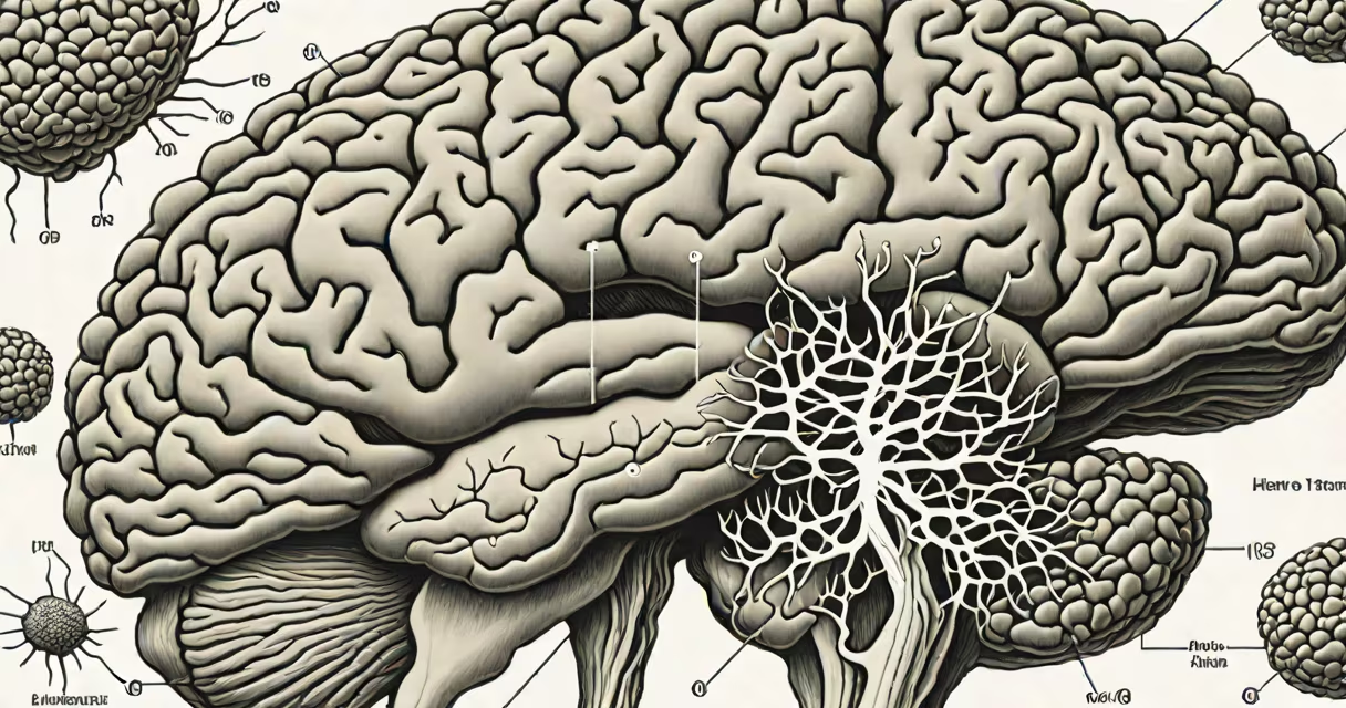
Prion disease in humans stems from abnormal folding of the prion protein (PrP), which accumulates in the brain and destroys neural tissue. These misfolded proteins are infectious and propagate by converting normal cellular prion proteins into their pathological form. The process leads to rapidly progressive dementia, motor dysfunction, and death.
Prions are unique among disease agents because they lack DNA or RNA. Instead, they are composed solely of misfolded proteins that replicate by inducing structural changes in healthy versions of the same protein. This mechanism defies the central dogma of molecular biology, where genetic material directs protein synthesis. Once initiated, the cascade of misfolding overwhelms the brain's clearance systems, resulting in neuronal loss and characteristic spongiform vacuolation visible under the microscope.
The human prion protein gene (PRNP) encodes a normal cellular protein (PrPC) that is harmless and abundant in the central nervous system. When it adopts an alternative conformation (PrPSc), it becomes resistant to degradation and aggregates into toxic fibrils. These aggregates disrupt cell function, trigger inflammation, and lead to synaptic failure and neuronal death. The disease progresses relentlessly, with survival typically measured in months after symptom onset.
There are four main categories of prion disease in humans, each differing in origin and clinical presentation. They are classified based on whether the condition arises spontaneously, genetically, or through exposure to infected material.
| Type | Origin | Prevalence | Examples |
|---|---|---|---|
| Sporadic | Unknown cause; random mutation | 85-90% | Sporadic Creutzfeldt-Jakob disease (sCJD) |
| Genetic | Inherited mutations in PRNP gene | 10-15% | Familial CJD, Gerstmann-Sträussler-Scheinker syndrome (GSS), Fatal Familial Insomnia (FFI) |
| Iatrogenic | Medical procedures involving contaminated instruments or tissues | Rare | Transmission via corneal transplants, dura mater grafts, growth hormone therapy |
| Acquired (variant) | Consumption of BSE-contaminated beef | Very rare | Variant CJD (vCJD) |
Sporadic CJD accounts for the majority of cases. It occurs without known risk factors and likely results from a spontaneous misfolding event in a single PrP molecule. The incidence is approximately 1 case per million people annually worldwide, according to the World Health Organization (WHO).
Genetic forms arise from autosomal dominant mutations in the PRNP gene. Over 20 such mutations have been identified, each associated with different disease phenotypes. For example, the D178N mutation causes either familial CJD or FFI depending on the codon 129 polymorphism (methionine or valine). Penetrance is high but age of onset varies widely, from early adulthood to late life.
Iatrogenic transmission has occurred through medical interventions using contaminated materials. A notable case involved patients receiving human-derived pituitary growth hormone extracted from cadaveric pituitaries between 1958 and 1985. Of those treated, 16 developed CJD, with incubation periods ranging from 5 to 20 years. The UK's National Health Service reported these cases in 2004, confirming the long latency period of iatrogenic prion disease.
Variant CJD emerged in the UK during the 1990s following the Bovine Spongiform Encephalopathy (BSE) epidemic. Unlike classical CJD, vCJD affects younger individuals (median age 28) and presents with prominent psychiatric symptoms and sensory disturbances before neurological decline. The UK's National Prion Monitoring Unit documented 228 confirmed cases by 2023, with the last death recorded in 2019.
Prion diseases are not contagious through casual contact. Transmission requires direct exposure to infected neural tissue or certain bodily fluids. Blood transfusion has been implicated in rare cases: one report described a patient who developed vCJD after receiving blood from a donor later diagnosed with the disease. The UK's National Blood Service suspended donations from individuals who had spent time in the UK during the BSE outbreak.
Environmental persistence is a key concern. Prions resist standard sterilization methods including autoclaving at 134°C for 18 minutes. They remain infectious on surgical instruments unless treated with sodium hydroxide or bleach solutions. The U.S. Centers for Disease Control and Prevention (CDC) recommends decontamination protocols involving 1N NaOH and extended heating times for instruments used in neurosurgery.
There is no evidence that prion disease spreads through saliva, sweat, or respiratory droplets. However, caution remains high in settings involving tissue handling. In 2018, a French study found prion protein in the urine of patients with sporadic CJD, though infectivity was not confirmed. This finding underscores the need for strict biosafety protocols in laboratories working with prion samples.
Many confuse prion disease with Alzheimer's or Parkinson's, both of which involve protein aggregation. However, unlike amyloid-beta or alpha-synuclein, prions are capable of self-replication and can transmit disease between individuals. The term "protein-only" reflects this distinction: no nucleic acid is required for propagation.
Another misconception is that prion disease is easily diagnosable in living patients. While MRI scans may show bilateral basal ganglia hyperintensity in 80% of CJD cases, and cerebrospinal fluid testing for 14-3-3 protein yields positive results in 70-80%, definitive diagnosis still requires postmortem brain examination. The sensitivity of CSF tests drops in variant CJD, where the pattern differs from classical CJD.
A non-obvious fact: the same prion strain can cause different diseases depending on host genetics. The PRNP codon 129 polymorphism determines susceptibility. Individuals homozygous for methionine (MM) are at higher risk for both sporadic and variant CJD. In contrast, valine/valine (VV) individuals are less susceptible. This explains why some populations show higher rates of disease despite similar exposure levels.
In 2004, a 39-year-old woman in the UK presented with progressive memory loss, visual disturbances, and myoclonus. Brain MRI revealed symmetric signal changes in the caudate nuclei. CSF analysis showed elevated 14-3-3 protein. She died six months later. Postmortem examination confirmed vCJD with widespread prion deposition and spongiform change.
Her medical history included consumption of beef products during the BSE outbreak. Genetic testing revealed MM homozygosity at codon 129. This combination-exposure to BSE-infected meat and a susceptible genotype-led to disease onset. The case exemplifies the interplay between environmental exposure and host genetics in acquired prion disease.
Understanding prion disease mechanisms is critical for public health policy. Surveillance systems like the UK's National Prion Monitoring Unit track cases and monitor potential new clusters. Early detection remains elusive, but research into biomarkers such as RT-QuIC (real-time quaking-induced conversion) shows promise for antemortem diagnosis.
For further insight into metabolic disruptions linked to gastrointestinal symptoms, explore the causes of flatulence in humans. While gas production involves microbial fermentation, prion disease involves a far more insidious process-self-replicating proteins that dismantle the brain.