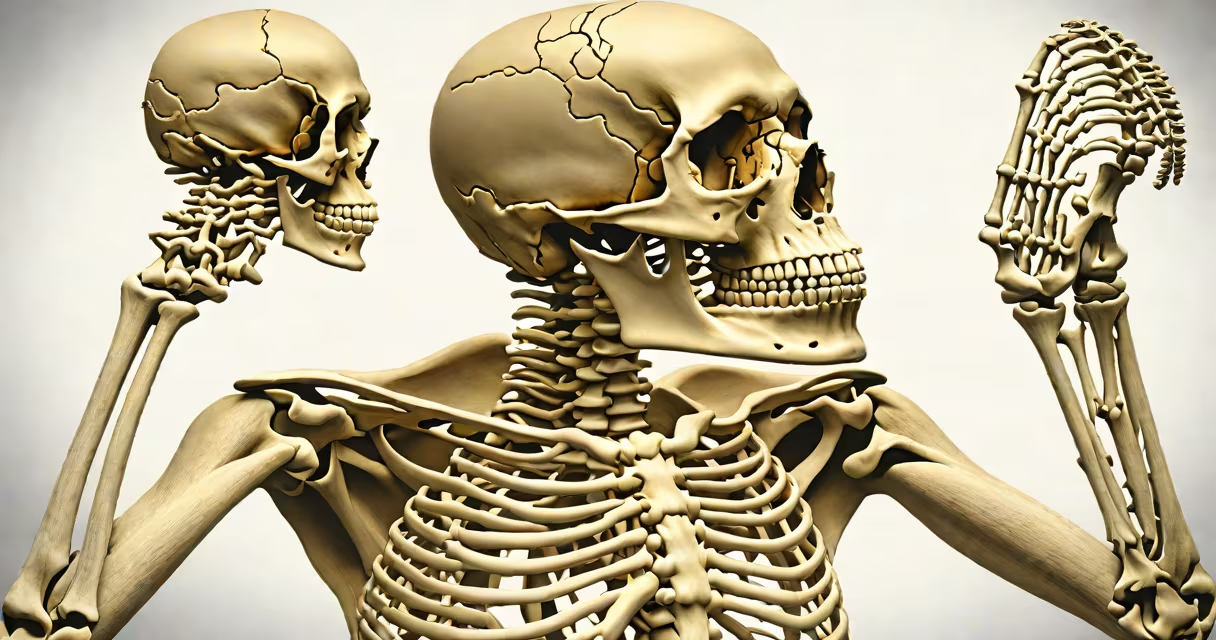
The study of skeletal system is the scientific examination of bones, joints, cartilage, and associated tissues that form the structure of the human body. It covers structure, function, growth, repair, and disease processes related to the skeleton. This field integrates anatomy, physiology, biomechanics, and pathology to explain how bones enable movement, protect important organs, and regulate calcium and phosphorus levels.
Skeletal biology focuses on the composition and behavior of bone tissue. Bones are living organs composed primarily of collagen fibers and hydroxyapatite crystals-a mineralized matrix that provides strength and flexibility. The human skeleton contains 206 bones in adults, distributed across the axial skeleton (skull, vertebral column, ribs, sternum) and appendicular skeleton (limbs, shoulder girdle, pelvic girdle).
Research shows that bone remodeling occurs continuously throughout life. Osteoclasts break down old bone, while osteoblasts build new bone. This process maintains structural integrity and regulates blood calcium levels. Disruptions in this cycle lead to conditions like osteoporosis, where bone density declines, increasing fracture risk. According to the National Institutes of Health, osteoporosis affects over 10 million Americans and contributes to more than 2 million fractures annually.
Scientists analyze the skeletal system through dissection, imaging techniques, and cellular assays. Radiographic methods such as X-ray, CT scans, and MRI allow visualization of bone structure and pathology without surgery. Microscopic analysis reveals the organization of bone layers-cortical (compact) bone and trabecular (spongy) bone-each serving distinct mechanical roles.
For example, cortical bone forms the outer shell of long bones and resists bending forces. Trabecular bone, found in vertebrae and the ends of long bones, acts like a shock absorber due to its porous network. A 2021 study published in *Bone Research* demonstrated that trabecular architecture correlates strongly with fracture resistance in older adults, especially in the femoral neck region.
Researchers also examine bone metabolism using biomarkers. Serum levels of C-terminal telopeptide (CTX) and procollagen type I N-terminal propeptide (P1NP) reflect bone resorption and formation rates, respectively. These markers help diagnose metabolic bone diseases and monitor treatment response in patients with osteoporosis.
Bone tissue is classified into four main types based on structure and location:
| Type | Structure | Location | Function |
|---|---|---|---|
| Cortical bone | Dense, compact layer | Shaft of long bones, skull | Support, protection, load-bearing |
| Trabecular bone | Porous, lattice-like | Vertebrae, hip, ends of long bones | Shock absorption, nutrient exchange |
| Woven bone | Random fiber alignment | Healing fractures, fetal development | Initial repair scaffold |
| Membranous bone | Formed via intramembranous ossification | Skull flat bones, clavicle | Direct bone formation without cartilage template |
One non-obvious fact: the skull's flat bones develop through intramembranous ossification, not endochondral ossification. This means they form directly from mesenchymal connective tissue membranes without a cartilage precursor. This distinction explains why certain skull fractures heal differently than long bone fractures.
During fetal development, most bones begin as cartilage models. Endochondral ossification replaces cartilage with bone in long bones like the femur. This process begins around week 6 of gestation and continues until early adulthood. Growth plates (epiphyseal plates) remain active until bone maturation, typically between ages 16 and 25.
Many assume bones are inert structures. In reality, they are metabolically active tissues. A single bone can contain over 100,000 blood vessels and thousands of nerves. Bone marrow within the medullary cavity produces red blood cells, platelets, and white blood cells-a function often overlooked.
Another misconception is that all joint movements involve bones sliding past each other. Synovial joints allow motion through fluid-filled cavities, but many bones are connected by fibrous or cartilaginous joints that limit or prevent movement. For example, the sutures in the skull are fibrous joints that fuse during adolescence, preventing movement to protect the brain.
Some confuse the skeletal system with the muscular system. While both contribute to movement, the skeletal system provides passive structure and use, whereas muscles generate force through contraction. The interaction between the two systems enables coordinated motion-such as walking or lifting-through a system called the musculoskeletal unit.
Understanding the study of skeletal system informs clinical practice. Orthopedic surgeons rely on detailed knowledge of bone structure to perform joint replacements. Total knee arthroplasty, for instance, requires precise alignment of prosthetic components to mimic natural joint mechanics and reduce wear over time.
A real-world example: In 2019, a patient underwent a complex reconstruction after a high-energy trauma involving a comminuted tibial fracture. Surgeons used CT-guided navigation and custom titanium implants based on 3D-printed models derived from preoperative scans. The procedure succeeded because the surgical team applied principles from bone structure skeletal system to restore weight-bearing stability.
On a broader scale, forensic anthropologists use skeletal remains to estimate age, sex, stature, and ancestry. The pubic symphysis and auricular surface of the ilium show predictable changes with age, allowing estimation within ±5 years in adults. These methods are standard in identifying victims of mass disasters or historical remains.
I once observed a cadaver lab session where students dissected the pelvis to identify the sacroiliac joints. The challenge wasn't just locating the articulation-it was understanding how its irregular shape and ligamentous reinforcement limit motion while transferring weight from the spine to the lower limbs. That hands-on experience clarified how form follows function in skeletal design.