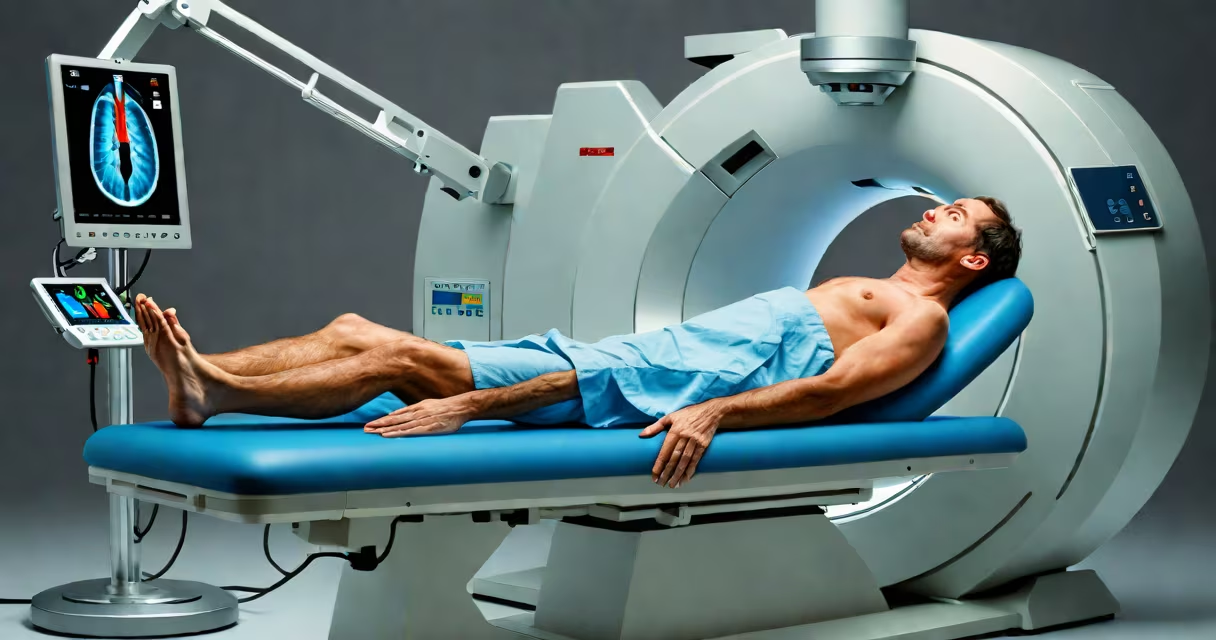
A ct scan procedure is a non-invasive medical imaging technique that uses rotating X-ray beams and computer reconstruction to produce detailed cross-sectional images of internal structures. It allows doctors to visualize bones, organs, blood vessels, and soft tissues with high accuracy, aiding in diagnosis and treatment planning.
The process begins with the patient lying on a motorized table that slides into a circular gantry. As the machine rotates around the body, it captures multiple X-ray images from different angles. A computer then combines these images into thin, slice-like views of the scanned area. These slices can be stacked to form 3D reconstructions, offering a complete view of anatomical details.
Most ct scan procedures take between 10 and 30 minutes, depending on the body region being imaged and whether contrast dye is used. The scan itself is painless, though patients must remain still to avoid blurring the images. Some scans require intravenous contrast to improve visibility of blood vessels or tumors.
During a ct scan procedure, the patient lies on a table that moves through a doughnut-shaped scanner. Inside the scanner, an X-ray tube rotates rapidly around the body, emitting narrow beams of radiation. On the opposite side, detectors measure how much radiation passes through each tissue type. Dense materials like bone absorb more radiation, while softer tissues allow more through.
The data collected by the detectors is sent to a computer, which applies complex mathematical algorithms-specifically, filtered back-projection-to reconstruct the raw data into cross-sectional images. Each image represents a single slice, typically 1 to 10 millimeters thick. Modern scanners can acquire hundreds of slices in under a minute.
For certain exams, such as abdominal or vascular scans, a contrast agent is injected into a vein. This dye highlights blood vessels and helps distinguish normal from abnormal tissue. The injection may cause a warm sensation or metallic taste, which resolves quickly. Patients are monitored during contrast administration for allergic reactions, though severe responses are rare.
CT scan procedures vary based on the body region and clinical purpose. The most common types include:
| Type | Body Region | Common Uses | Typical Scan Time |
|---|---|---|---|
| Head CT | Brain and skull | Stroke detection, trauma evaluation, tumor identification | 5-10 minutes |
| Abdominal CT | Abdomen and pelvis | Appendicitis, kidney stones, cancer staging | 10-20 minutes |
| Chest CT | Lungs and heart | Pulmonary embolism, lung nodules, aortic dissection | 10-15 minutes |
| CT Angiography (CTA) | Blood vessels | Coronary artery disease, aneurysms, vascular malformations | 15-25 minutes |
Each type targets specific conditions. For example, a head CT scan can detect hemorrhage within minutes after a traumatic brain injury, guiding immediate surgical decisions. According to the American College of Radiology, head CTs are used in over 60% of emergency department evaluations for suspected stroke.
Contrast-enhanced scans provide higher sensitivity for detecting small lesions. A study published in *Radiology* found that contrast-enhanced abdominal CT improved detection of pancreatic tumors by 34% compared to non-contrast scans.
Patients are asked to remove metal objects, including jewelry and belts, because metal can distort the image. They wear a hospital gown and lie on the scanning table, usually on their back. The technologist positions the patient and may place cushions or straps to maintain alignment.
Once inside the scanner, the patient hears a whirring sound as the machine rotates. The technician monitors from a separate room via a window and intercom. Communication is possible throughout. The patient must hold their breath briefly during some scans-especially chest or abdominal-to prevent motion blur.
After the scan, the patient can resume normal activities immediately unless sedation was used. Results are interpreted by a radiologist, who sends a report to the referring physician within 24 hours. In urgent cases, such as trauma or suspected stroke, results may be available in under an hour.
I once observed a head ct scan procedure on a patient with sudden onset aphasia. The scan revealed a large left middle cerebral artery infarction. The neurology team initiated thrombolytic therapy within 45 minutes of arrival, a decision supported by the rapid imaging.
One widespread misunderstanding is that ct scan procedures involve high radiation exposure. While they do use ionizing radiation, the dose varies by type. A standard head CT delivers about 2 millisieverts (mSv), comparable to 8 months of natural background radiation. A full-body CT may reach 10 mSv, still below the annual limit recommended by the National Council on Radiation Protection and Measurements (NCRP).
Another confusion is equating ct scans with MRIs. Both generate cross-sectional images, but they use different technologies. CT relies on X-rays and is faster and better for bone and acute bleeding. MRI uses magnetic fields and radio waves, excels in soft tissue detail, and avoids radiation-but takes longer and is contraindicated in patients with certain implants.
Some assume ct scan procedures are painful. They are not. The machine does not touch the body. The only discomfort may come from lying still or receiving an IV contrast injection, which feels like a brief warmth in the chest or face.
Doctors order ct scan procedures when quick, accurate visualization of internal structures is needed. Common reasons include evaluating trauma, diagnosing infections, staging cancer, and assessing vascular health. In emergency medicine, ct scans are first-line tools for head injuries and suspected strokes.
For example, a patient presenting with severe headache and neurological deficits may undergo a non-contrast head CT to rule out hemorrhage. If negative, a follow-up MRI may be ordered for ischemic stroke. The CT scan provides rapid triage, enabling timely interventions.
According to the U.S. Food and Drug Administration, over 80 million ct scans are performed annually in the United States. The majority are for oncology, trauma, and cardiovascular assessment.