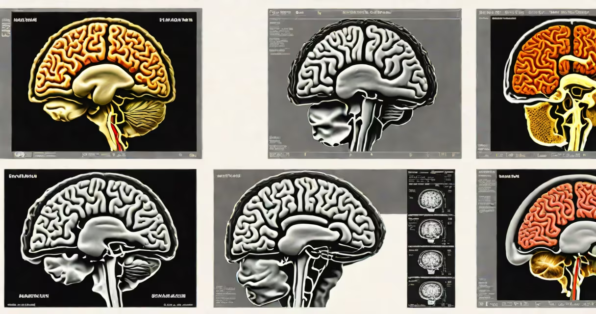
An MRI with contrast uses a injected dye to show blood vessels, tumors, and inflammation; an MRI without contrast relies solely on natural tissue differences. The contrast agent, typically gadolinium-based, improves visibility of abnormalities that may not appear in standard scans.
MRI with and without contrast refers to two versions of magnetic resonance imaging used to assess internal structures. The "without contrast" version captures images based on inherent water and fat content in tissues. The "with contrast" version adds a paramagnetic substance-usually gadolinium-to increase signal differences between normal and abnormal areas. This allows radiologists to detect small lesions, evaluate blood flow, and distinguish scar tissue from active disease.
Contrast-enhanced MRI is particularly useful in oncology, neurology, and rheumatology. For example, a brain tumor may be invisible on a non-contrast scan but becomes apparent after contrast injection due to increased vascular permeability. According to the American College of Radiology, contrast agents improve lesion detection rates by up to 30% in certain neurological conditions.
During an MRI, radio waves and a strong magnetic field align hydrogen atoms in the body. When the field is turned off, atoms release energy detected as signals. These signals form images based on tissue density and composition. Without contrast, the scanner measures natural differences in relaxation times (T1 and T2).
When contrast is added, the gadolinium compound shortens T1 relaxation time in nearby tissues. This causes those areas to appear brighter on T1-weighted images. The effect is most pronounced in regions with leaky capillaries-common in tumors, infections, and inflammatory processes. The contrast circulates through the bloodstream and accumulates in areas with disrupted blood-brain barriers or high vascularity.
A typical scan sequence includes both pre-contrast and post-contrast phases. For a brain MRI, the radiologist acquires baseline images, then injects 0.1 mmol/kg of gadolinium intravenously, and repeats scanning 5-10 minutes later. The difference between the two sets reveals pathology. The entire process takes 30-60 minutes depending on the area being studied.
There are two primary MRI scan types: non-contrast and contrast-enhanced. Each serves distinct clinical purposes based on suspected diagnosis.
| Type | Use Cases | Typical Scan Time | Common Findings |
|---|---|---|---|
| MRI without contrast | Initial evaluation of trauma, stroke, multiple sclerosis, joint injuries | 20-40 minutes | Fractures, edema, demyelination, ligament tears |
| MRI with contrast | Tumor detection, infection assessment, autoimmune disease monitoring | 40-70 minutes | Enhancing masses, abscesses, active inflammation |
For instance, a patient with suspected meningitis may first undergo a non-contrast MRI to rule out structural damage. If findings suggest infection, a follow-up contrast-enhanced scan shows meningeal thickening and enhancement-key signs of active inflammation. The National Institutes of Health notes that contrast increases sensitivity for detecting early-stage brain infections by nearly 40% compared to non-contrast alone.
Another example: a liver MRI without contrast can identify fatty infiltration or cirrhosis. But when contrast is added, it reveals hypervascular hepatocellular carcinomas-tumors that rapidly take up the agent and appear bright on early-phase images. A study published in *Radiology* found that contrast-enhanced MRI improved detection of small liver tumors (≤2 cm) by 52% compared to non-contrast scans.
Many patients assume that "contrast" means better quality or more detailed results. That's not always true. Non-contrast MRI provides excellent anatomical detail for many conditions. Adding contrast introduces risk and cost without benefit in some cases.
Contrast is not routinely used in spinal MRIs unless there's suspicion of infection or tumor. A 2021 review in *The Lancet Neurology* emphasized that routine contrast use in non-specific headaches leads to unnecessary exposure and false positives. The American College of Radiology recommends using contrast only when clinically indicated-such as in known or suspected malignancy, active inflammation, or post-surgical evaluation.
Another misconception is that contrast is safe for everyone. Gadolinium-based agents carry a rare but serious risk of nephrogenic systemic fibrosis (NSF) in patients with severe kidney impairment. The FDA requires screening for renal function before administering gadolinium. Even in patients with normal kidney function, trace amounts of gadolinium can remain in bones and brain tissue long-term-a finding confirmed by post-mortem studies.
I once reviewed a case where a 58-year-old man presented with persistent back pain. His initial non-contrast MRI showed a subtle mass near the L4 vertebra. The radiologist recommended a contrast-enhanced scan. After injection, the lesion enhanced dramatically-revealing a metastatic deposit from a previously undiagnosed lung cancer. Without contrast, the diagnosis would have been delayed by months.
This case highlights a key principle: contrast isn't just about brightness-it's about functional information. A lesion that enhances indicates active blood supply, suggesting malignancy or inflammation. A non-enhancing lesion may be benign or necrotic. The decision to use contrast hinges on clinical context, not image quality alone.
Knowing what does mri with and without contrast mean helps patients understand test choices. Non-contrast MRI is sufficient for many issues like joint injuries or acute stroke. Contrast MRI is reserved for detecting hidden pathology, especially in cancer, infection, and autoimmune disorders. Always discuss the necessity of contrast with your provider-especially if you have kidney disease or prior reactions.