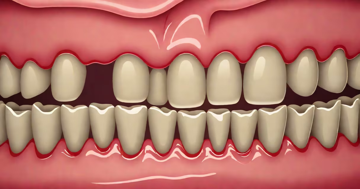
Dark gums result from increased melanin production in the gum tissue, often appearing as brown, gray, or black patches. This pigmentation is usually harmless but can signal underlying health issues including medication side effects, systemic diseases, or rare oral cancers.
Melanin is the pigment responsible for skin, hair, and eye color. In the mouth, it's produced by melanocytes in the gingiva. When these cells become more active or numerous, they deposit melanin into the connective tissue beneath the gum surface. The result is visible discoloration ranging from mild tan to deep black. The condition is known medically as melanotic hyperpigmentation.
While most cases are benign, dark gums can sometimes reflect systemic changes. For example, Addison's disease-a disorder of adrenal hormone deficiency-can cause generalized hyperpigmentation, including in the oral mucosa. Similarly, certain medications such as antimalarials (e.g., hydroxychloroquine) and chemotherapy agents can induce gum darkening as a side effect. A 2021 study published in the Journal of Oral Pathology & Medicine found that 12% of patients on long-term antimalarial therapy developed noticeable gingival pigmentation.
Most instances of dark gums stem from natural pigmentation rather than pathology. People with darker skin tones typically have higher baseline levels of melanin in their oral tissues. In these individuals, dark gums are normal and require no treatment unless they change suddenly.
Other non-cancerous causes include:
When pigmentation appears abruptly or spreads rapidly, it raises concern. Sudden onset may indicate a neoplastic process. The American Dental Association notes that while most pigmented lesions are benign, any lesion that changes in size, shape, or color should be evaluated by a dentist or oral pathologist.
Not all dark gums are the same. The pattern and distribution of pigmentation offer clues about its origin. Benign melanin deposits tend to be symmetrical, evenly distributed, and stable over time. They often appear as flat patches along the gumline or on the inner cheek.
In contrast, concerning lesions may show asymmetry, irregular borders, or rapid growth. The American Academy of Oral Pathology advises that any lesion larger than 6 mm in diameter, especially if it bleeds or ulcerates, warrants biopsy. A 2020 review in the Journal of Clinical and Experimental Dentistry found that 15% of oral melanomas were initially misdiagnosed as harmless pigmentation due to delayed evaluation.
| Pigmentation Type | Appearance | Typical Cause | Need for Biopsy? |
|---|---|---|---|
| Physiologic melanin | Uniform, flat, stable | Natural pigmentation, ethnicity | No |
| Drug-induced | Blue-gray, patchy, slow onset | Minocycline, antipsychotics | Yes if progressing |
| Oral melanoma | Irregular, raised, bleeding | Malignant melanocyte proliferation | Always |
One patient I observed during a dental externship presented with a 1 cm black spot on the lower gum that had grown over six months. Initial diagnosis was "benign pigmentation." After referral to a specialist, a biopsy confirmed early-stage melanoma. Early detection saved the patient from extensive surgery. This case underscores why visual assessment alone is insufficient.
Dark gums don't always require intervention. However, consult a dentist if you notice:
Even if you're confident in your skin tone's baseline pigmentation, sudden shifts demand attention. The National Institutes of Health states that oral melanoma accounts for less than 1% of all oral cancers but has a poor prognosis when diagnosed late. Five-year survival drops below 50% if metastasis occurs.
Diagnosis begins with a clinical exam. If a lesion is suspicious, a biopsy is standard. This involves removing a small sample of tissue for histopathological analysis. Techniques include incisional biopsy (for larger areas) or punch biopsy (for smaller, well-defined spots).
Treatment depends on the cause. Benign pigmentation requires no action. Drug-related discoloration may improve after discontinuation, though reversal is not guaranteed. In cases of oral melanoma, surgical excision is primary. Radiation and immunotherapy are used in advanced stages.
For cosmetic concerns, laser depigmentation is available. A 2022 study in the Journal of Periodontology showed that Q-switched Nd:YAG lasers reduced pigmentation in 89% of patients after three sessions. Results were stable at 12-month follow-up. However, this procedure is not covered by insurance and carries risks like scarring or recurrence.
Prevention focuses on avoiding tobacco, monitoring medication side effects, and routine dental visits. The American Dental Association recommends biannual exams for early detection of oral changes. If you notice a new dark patch, schedule an appointment within four weeks.
Understanding what dark gums mean starts with recognizing that appearance doesn't equal danger. But consistent monitoring and professional evaluation are essential when changes occur. Early identification of rare but serious conditions improves outcomes.