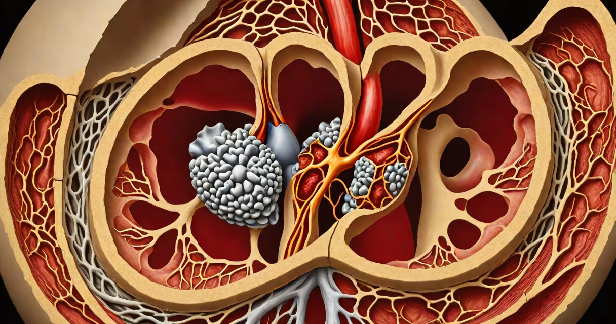
The aortic valve is a semilunar valve located between the left ventricle and the ascending aorta, regulating blood flow from the heart into the systemic circulation. It opens during systole to allow oxygenated blood to exit the left ventricle and closes during diastole to prevent backflow.
Composed of three thin, flexible cusps-left, right, and posterior-this valve functions as a one-way gate. Each cusp is shaped like a half-moon, hence the term "semilunar." During ventricular contraction, pressure builds in the left ventricle, pushing the cusps apart. As pressure drops after contraction, the cusps snap shut due to elastic recoil and blood backpressure.
Normal aortic valve function depends on precise geometry and tissue elasticity. The valve sits at the level of the aortic root, where the aorta begins its ascent from the heart. Its position ensures efficient unidirectional flow and prevents volume overload on the left ventricle.
When the left ventricle contracts, intraventricular pressure exceeds aortic pressure. This pressure gradient forces the three cusps to open outward, creating a central orifice through which blood flows into the aorta. The opening diameter reaches approximately 2.5 cm in adults during peak systole.
As ventricular relaxation begins, aortic pressure exceeds ventricular pressure. Blood starts to flow backward toward the ventricle. The cusps close rapidly due to their inherent elasticity and the force of returning blood. Closure generates the second heart sound (S2), audible with a stethoscope at the base of the heart.
Valve closure timing is critical. Delayed closure leads to regurgitation-blood flowing back into the ventricle. This increases workload on the heart and can cause left ventricular hypertrophy over time. Prolonged regurgitation may progress to heart failure if untreated.
The aortic valve is composed of fibrous connective tissue rich in collagen and elastin. These materials provide strength and flexibility. The cusps are anchored to the aortic annulus-a ring of fibrous tissue that forms the valve's base.
Each cusp has a central fibrosa layer, a thickened region that resists stretching under high pressure. The free edges of the cusps are thin and avascular, relying on diffusion from surrounding tissues for nutrition. This makes them vulnerable to calcification over time.
Unlike atrioventricular valves (like the mitral valve), the aortic valve lacks chordae tendineae. Instead, it relies on passive hemodynamic forces for closure. The absence of tethering structures allows for rapid, low-resistance opening and sealing.
| Feature | Description |
|---|---|
| Number of cusps | Three (left, right, posterior) |
| Shape | Semi-lunar (crescent-shaped) |
| Location | Between left ventricle and ascending aorta |
| Opening pressure | ~30 mmHg (during systole) |
| Closure mechanism | Hemodynamic backpressure and elastic recoil |
Many assume the aortic valve is a single flap. It is not. It has three cusps, unlike the mitral valve, which has two. This tri-leaflet design allows for uniform stress distribution and efficient closure under high-pressure conditions.
Another misconception is that the valve opens passively. It does not. Opening requires sufficient ventricular pressure-approximately 30 mmHg-to overcome the resistance of the aortic wall and the valve's own stiffness. Without adequate contractility, the valve remains closed, limiting perfusion.
A less obvious point: the aortic valve is not symmetric. The right cusp is largest and most mobile. The left cusp is smallest and often fused with the anterior wall of the aortic root. This asymmetry influences surgical repair techniques and prosthetic valve placement.
Calcific aortic stenosis occurs when calcium deposits build on the cusps, reducing mobility. This condition affects 2% of adults over 65 and is the most common cause of aortic valve replacement. A patient with severe stenosis may have a valve orifice area below 1.0 cm², leading to symptoms like syncope, angina, and dyspnea.
For example, a 78-year-old man presented with exertional chest pain and fainting. Echocardiography revealed a peak transvalvular gradient of 75 mmHg and a valve area of 0.8 cm²-diagnostic of severe aortic stenosis. He underwent transcatheter aortic valve implantation (TAVI), a minimally invasive procedure replacing the native valve with a prosthetic one.
Another clinical scenario involves infective endocarditis. Bacteria such as *Streptococcus viridans* can colonize damaged cusps, forming vegetations. These can embolize to the brain or kidneys. Early diagnosis via transesophageal echocardiography is important-delayed treatment increases mortality risk.
I once observed a live cardiac surgery where a surgeon removed a heavily calcified aortic valve and implanted a bioprosthetic valve. The patient had a history of rheumatic fever, which had led to scarring and reduced cusp motion. The surgery restored normal flow dynamics, confirmed by intraoperative Doppler ultrasound.
The aortic valve works in tandem with the aortic root and the coronary arteries. The left and right coronary arteries originate from the sinuses of Valsalva-small pockets in the aortic wall just above the valve. These sinuses expand during systole, allowing blood to enter the coronary vessels and supply the myocardium.
Disruption of this relationship can impair coronary perfusion. For instance, in Marfan syndrome, abnormal connective tissue weakens the aortic wall, leading to dilation of the root and eventual valve incompetence. This condition increases the risk of aortic dissection and sudden death.
Understanding the aortic valve anatomy is essential for interpreting imaging studies. Transthoracic echocardiography (TTE) provides a standard view of the valve, while transesophageal echocardiography (TEE) offers higher resolution, especially useful in detecting small vegetations or subtle regurgitation.
aortic valve anatomy defines the physical and functional basis of one of the heart's most important components. Its three cusps, precise positioning, and reliance on hemodynamic forces ensure efficient blood ejection and prevent backflow. Understanding this structure supports accurate diagnosis, effective treatment, and better outcomes in patients with valvular disease.